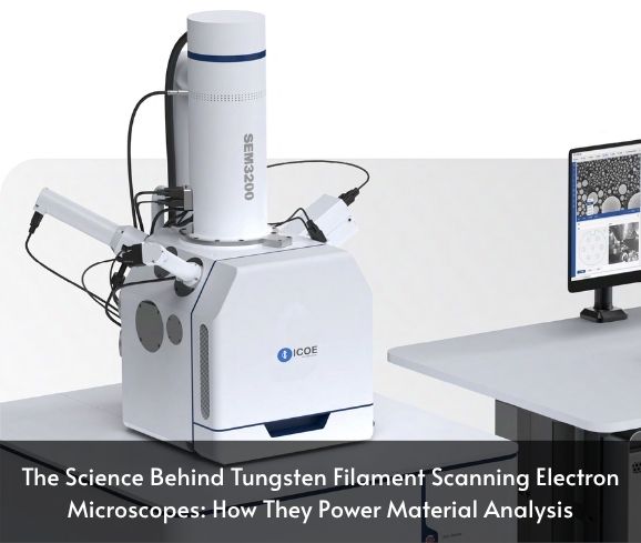
The Basic Physics of the Tungsten Filament Scanning Electron Microscope
Understanding the tungsten filament scanning electron microscope starts with its core scientific principle—thermionic emission. In these systems, a tungsten filament is heated to extremely high temperatures, typically over 2700°C. At this temperature, the tungsten wire begins to emit electrons. These electrons are then accelerated toward the sample using a series of electric fields in a vacuum chamber.
The accelerated electrons strike the surface of the sample, causing a cascade of interactions, including the emission of secondary electrons. These secondary electrons are captured by detectors, allowing the microscope to generate highly detailed surface images. The tungsten filament scanning electron microscope works particularly well for examining topographies, morphologies, and microstructural features of a wide range of materials.
At Hansvue, we recognize how this foundational science translates into practical benefits for labs and industries in Malaysia and beyond.
Advantages of a Tungsten Filament Scanning Electron Microscope in Research
There are numerous reasons why the tungsten filament scanning electron microscope is widely used in scientific research and academic institutions.
1. Cost-Effective Entry to SEM Technology
For researchers and educational labs operating under budget constraints, the tungsten filament scanning electron microscope provides the best value. Compared to field emission and LaB6 SEMs, it is significantly more affordable, both for initial investment and operational costs.
2. Ease of Maintenance
Tungsten filaments are easy to replace, making these systems simple to maintain. At Hansvue, our SEM units are designed with accessible filament compartments and user-friendly maintenance guidelines.
3. Reliable for General Imaging
For most material science applications—including failure analysis, particle inspection, and crystallographic imaging—the tungsten filament scanning electron microscope offers reliable results. It may not reach the ultra-high resolution of field emission SEMs, but it provides excellent clarity for general imaging needs.
4. Educational Utility
In universities, the tungsten filament scanning electron microscope serves as a practical tool to train students in SEM operation without the risks or costs of more complex systems.
Hansvue offers support and training packages to ensure researchers and students maximize their microscope’s potential.
Key Components of a Tungsten Filament Scanning Electron Microscope
A well-engineered tungsten filament scanning electron microscope is composed of several key components that work together to deliver clear and consistent imaging:
1. Electron Gun
This houses the tungsten filament and is responsible for generating the electron beam. Tungsten is chosen for its high melting point and stable thermionic emission properties.
2. Condenser and Objective Lenses
These electromagnetic lenses focus the beam into a fine point, enhancing resolution and image accuracy.
3. Scanning Coils
The beam is rastered over the surface of the specimen using scanning coils, ensuring full coverage and precision.
4. Sample Chamber
Samples are placed in a vacuum environment, allowing electrons to interact with the sample surface without air interference.
5. Electron Detectors
Secondary and backscattered electron detectors gather signals to generate detailed images. Some tungsten filament scanning electron microscopes also support EDS (Energy Dispersive X-ray Spectroscopy) for elemental analysis.
6. Control System & Software
Hansvue SEMs come equipped with intuitive control software to handle scanning parameters, imaging adjustments, and data exportation.
Each component in Hansvue’s tungsten filament scanning electron microscope lineup is selected and calibrated for optimal synergy, ensuring reliable results across every scan.
Enhancing Imaging Capabilities with a Tungsten Filament Scanning Electron Microscope
Modern research demands increasingly detailed imagery, and Hansvue understands that imaging quality is crucial—even in cost-effective systems. That’s why our tungsten filament scanning electron microscopes come with features that enhance overall imaging performance.
1. High Stability Design
Hansvue SEMs minimize vibration and electrical interference, allowing for steady imaging even at high magnifications.
2. Variable Pressure Modes
Some of our tungsten filament scanning electron microscope models support low-vacuum or variable pressure imaging, enabling researchers to examine non-conductive or hydrated samples without extensive coating.
3. Optimized Electron Optics
Hansvue systems incorporate refined electromagnetic lenses to maintain beam coherence and reduce aberrations, enhancing image clarity.
4. Smart Automation
Automation features like auto-focus, auto-brightness, and sample navigation make our tungsten filament scanning electron microscope systems more user-friendly—ideal for busy research environments.
Why the Tungsten Filament Scanning Electron Microscope Remains Relevant
Despite rapid advancements in microscopy, the tungsten filament scanning electron microscope continues to hold a significant place in laboratories and industries.
1. Simplicity Meets Performance
Not every application requires ultra-high resolution or nanometer-scale imaging. For many users, the tungsten filament scanning electron microscope provides the right balance of power and simplicity.
2. Widespread Accessibility
Hansvue ensures that researchers across Malaysia—from Klang Valley to Penang and Johor—can access high-quality SEMs without prohibitive costs.
3. Versatile Applications
From academic teaching labs to industrial QA departments, the tungsten filament scanning electron microscope proves its versatility again and again.
This relevance is exactly why Hansvue maintains a strong inventory of tungsten filament SEMs and continues to invest in SEM support, training, and upgrades.
User Experiences with Tungsten Filament Scanning Electron Microscopes
Hansvue’s clients range from research institutions and universities to industrial manufacturers. Here’s what some users have reported after incorporating a tungsten filament scanning electron microscope into their workflow:
University of Malaya: “Our lab relies on the Hansvue SEM for undergraduate teaching. It’s easy to use, durable, and delivers high-quality images that rival more expensive systems.”
Medical Device Manufacturer in Selangor: “The SEM helps us monitor surface defects in titanium implants with precision. The tungsten filament model is both cost-effective and efficient.”
Materials Lab in Penang: “Switching to Hansvue’s tungsten filament scanning electron microscope improved our imaging throughput and allowed us to meet ISO inspection standards.”
These real-world results highlight the effectiveness and reliability of the tungsten filament scanning electron microscope in diverse fields.
Hansvue’s Expertise in Tungsten Filament Scanning Electron Microscope Solutions
At Hansvue, we specialize in delivering tailored microscopy solutions across Southeast Asia. Our tungsten filament scanning electron microscopes are selected for their durability, performance, and cost-efficiency.
What Sets Hansvue Apart?
Technical Knowledge: Our team includes SEM specialists who understand your unique imaging needs.
Installation & Training: We don’t just deliver your microscope—we ensure your team knows how to use it.
After-Sales Support: Whether it’s filament replacement, software updates, or image troubleshooting, Hansvue is just a call away.
Customization: We offer upgrades and accessories like sample holders, EDS detectors, and vibration-isolation tables.
With Hansvue, your tungsten filament scanning electron microscope is not just a purchase—it’s an investment in quality and service.
Conclusion
The tungsten filament scanning electron microscope represents a proven, accessible, and versatile solution for material analysis. Whether you’re in academic research, product development, or quality assurance, this SEM technology offers high-quality imaging without the high cost.
Hansvue remains committed to empowering Malaysian and Southeast Asian researchers with dependable tungsten filament scanning electron microscopes. Backed by expert support, training, and long-term maintenance, our SEM solutions are ready to meet your lab’s imaging demands—today and tomorrow.